创新背景
超声成像利用超声声束扫描人体,接受处理反射信号以获得体内器官的图象。超声图像可以为临床医生提供患者内脏器官的实时图像,安全无创地了解人体运作。但目前超声成像需要笨重的专用设备,只有医院和医生办公室才能使用。
创新过程
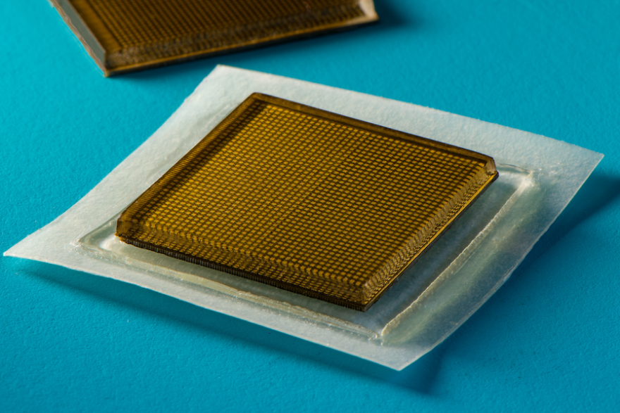
麻省理工学院工程师设计了一种新的超声成像设备,是一种大小类似邮票的超声波贴纸,可以附着在皮肤上提供48小时内内部器官的连续超声成像,使超声成像技术可以像在药房购买创可贴一样可穿戴且易于使用。
新设备将弹性粘合剂层与刚性传感器阵列配对,以便在更长时间内产生高分辨率的图像。粘合层由薄薄的两篇弹性体组成,弹性体中封装了容易传递声波的水基材料固体水凝胶,弹性体可以防止水凝胶脱水,水凝胶高度水合时声波能够有效穿透,提供内部器官的高分辨率成像。

粘合层与传感器组合使设备能够贴合皮肤,同时保持传感器的相对位置。底部弹性体层设计使设备能够粘附在皮肤上,顶层则粘附在团队设计和制造的刚性传感器阵列上。
研究人员将贴纸运用于志愿者来进行一系列测试。技术人员首先将液体凝胶涂在患者的皮肤上传输超声波,然后将探针或换能器压在凝胶上,将声波发送到体内,这些声波从内部结构回声并返回到探针,在那里回波信号被转换为视觉图像。志愿者将贴纸贴在身体的各个部位,附着在皮肤上。

液体超声凝胶会随着时间的流逝而流走并变干,中断长期成像。研究人员花了数年时间探索可拉伸超声探头的设计。探头将为内部器官提供便携式、低调的成像,提供了一系列灵活的微型超声换能器,可以随着患者身体移动伸展,产生的图像分辨率较低。
超声贴纸附着在志愿者皮肤上,长达48小时持续产生主要血管和更深层器官的清晰图像,且志愿者再次期间在实验室可以自由活动做各种动作。研究员从贴纸的图像中观察到坐着和站立时主要血管直径的变化。贴纸还捕捉了心脏在运动过程中改变形状的过程。
研究人员通过贴纸的超声成像功能观察胃膨胀,发现它在志愿者喝酒时收缩,然后从他们的系统中排出果汁。当一些志愿者举重时,贴纸检测到下层肌肉的明亮模式,发出暂时性微损伤的信号。研究人员表示,通过成像或许可以在过度使用肌肉之前捕捉锻炼中的时刻,为专家提供可以解释的成像数据。
目前的设计需要将贴纸连接到将反射声波转换为图像的仪器上,类似心脏监测EKG贴纸可以应用于医院的患者,并且可以连续成像内部器官,无需技术人员长时间将探头固定到位。
团队目前的目标是将超声波贴纸制成可穿戴成像产品,满足无线操作的需求,患者可以将其从医生办公室带回家或在药房购买。研究将进一步开发基于人工智能的软件算法,更好地解释和诊断贴纸的图像,患者和消费者可以购买贴纸监测内脏器官和肿瘤情况以及胎儿的发育。
创新关键点
结合弹性粘合剂和刚性传感器制作超声丞相贴纸,弹性体延长贴纸监测时间,为医疗成像技术拓展新方法。
创新价值
创新超声成像的设备,将工学技术与医学需求相结合,突破现有医学成像技术和可穿戴设备。
Innovative ultrasonic imaging equipment for elastomer-paired sensors
MIT engineers have designed a new ultrasound imaging device, an ultrasound sticker the size of a stamp that can be attached to the skin to provide continuous ultrasound imaging of internal organs over a 48-hour period, making ultrasound imaging technology as wearable and easy to use as buying a Band-Aid in a pharmacy.
The new device pairs the elastic adhesive layer with a rigid sensor array to produce high-resolution images over a longer period of time. The adhesive layer consists of a thin two-piece elastomer encapsulated in a solid hydrogel, a water-based material that can easily transmit sound waves, which prevents hydrogel dehydration, and sound waves can penetrate effectively when the hydrogel is highly hydrated, providing high-resolution imaging of internal organs.
The adhesive layer combined with the sensor allows the device to fit the skin while maintaining the relative position of the sensor. The bottom elastomer layer design allows the device to adhere to the skin, while the top layer adheres to the rigid sensor array designed and manufactured by the team.
The researchers used stickers on volunteers to conduct a series of tests. The technician first applies a liquid gel to the patient's skin to transmit ultrasound, then presses a probe or transducer onto the gel to send sound waves into the body, where these sound waves echo from the internal structure and back to the probe, where the echo signal is converted into a visual image. Volunteers attach stickers to various parts of the body and attach them to the skin.
Liquid ultrasound gels flow away and dry out over time, interrupting long-term imaging. Researchers have spent years exploring the design of stretchable ultrasound probes. The probe will provide portable, low-profile imaging of internal organs, providing a range of flexible miniature ultrasound transducers that can be extended as the patient's body moves, resulting in low-resolution images.
Ultrasound stickers attached to the volunteers' skin for up to 48 hours continued to produce clear images of major blood vessels and deeper organs, and the volunteers were free to do various movements in the lab again. The researchers observed changes in the diameter of major blood vessels when sitting and standing from images of stickers. Stickers also capture the process by which the heart changes shape during exercise.
The researchers observed stomach bloating through the sticker's ultrasound imaging function and found that it contracted as the volunteers drank alcohol and then expelled juice from their systems. When some volunteers lifted weights, the sticker detected bright patterns in the underlying muscles, signaling temporary micro-injuries. The researchers say imaging may be possible to capture moments during exercise before overusing muscles, providing experts with interpretable imaging data.
Current designs require attaching stickers to instruments that convert reflected sound waves into images, and similar to heart-monitoring EKG stickers can be applied to patients in hospitals and can continuously image internal organs without the need for technicians to hold the probe in place for long periods of time.
The team's current goal is to make ultrasound stickers into wearable imaging products that meet the demands of wireless operations that patients can take home from a doctor's office or purchase at a pharmacy. The research will further develop AI-based software algorithms to better interpret and diagnose images of stickers that patients and consumers can purchase to monitor internal organs and tumor conditions as well as fetal development.
智能推荐
遥感技术创新思维 | 创新开发微型开关可提供固态 LiDAR 记录分辨率
2022-11-08研究人员开发了新型高分辨率激光雷达芯片,可提供固态的 LiDAR 记录分辨率。
涉及学科涉及领域研究方向