创新背景
通过脑部扫描来寻找动脉瘤的迹象意味着要浏览数百张图像。动脉瘤有很多种大小和形状,并以复杂的角度膨胀——有些仅仅是一连串电影图像中的一个小光点。寻找动脉瘤是放射科医生最劳动密集和最关键的任务之一。
创新过程
这个工具是围绕一个叫做HeadXNet的算法构建的,它提高了临床医生正确识别动脉瘤的能力,其水平相当于在100次包含动脉瘤的扫描中发现6个以上的动脉瘤。它还提高了临床医生之间的共识。虽然HeadXNet在这些实验中的成功是有希望的,但研究团队——他们在机器学习、放射学和神经外科方面拥有专业知识——警告说,考虑到不同医院中心的扫描仪硬件和成像协议的差异,在实时临床部署之前,需要进一步的研究来评估AI工具的通用性。研究人员计划通过多中心合作来解决这些问题。
Yeom把这个想法带到了斯坦福大学机器学习小组(Machine Learning Group)举办的人工智能医疗保健训练营(AI for Healthcare Bootcamp),该小组由计算机科学兼职教授、该论文的资深合著者吴恩达(Andrew Ng)领导。主要的挑战是创建一个人工智能工具,可以准确地处理这些大量的3D图像,并补充临床诊断实践。

为了训练他们的算法,Yeom与Park和计算机科学研究生Christopher Chute合作,并在611次计算机断层扫描(CT)血管造影头部扫描中发现了临床重要的动脉瘤。
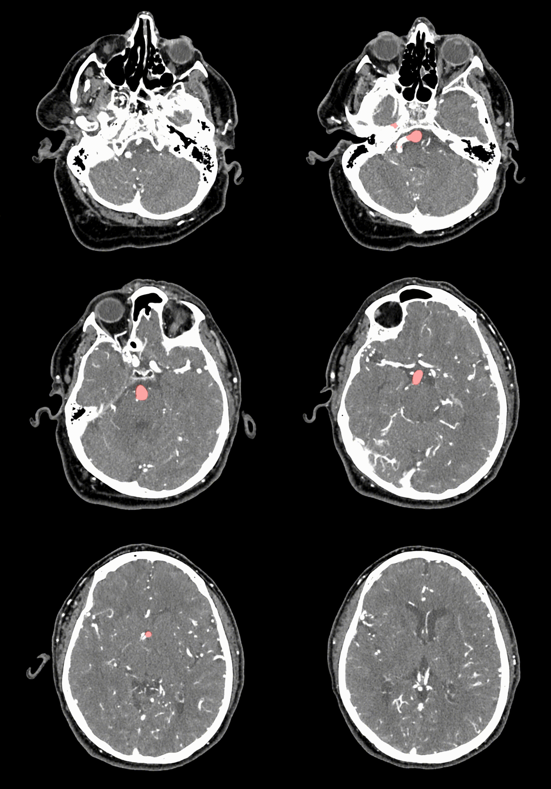
在训练之后,算法决定扫描的每个体素是否存在动脉瘤。HeadXNet工具的最终结果是算法的结论以半透明的高光覆盖在扫描上方。这种算法决策的表示使得临床医生很容易在没有HeadXNet输入的情况下仍能看到扫描结果。
8名临床医生对HeadXNet进行了测试,评估了115个动脉瘤的脑部扫描,一次有HeadXNet的帮助,一次没有。有了这个工具,临床医生正确地识别出更多的动脉瘤,因此减少了“漏诊”率,临床医生更有可能彼此达成一致意见。HeadXNet并不影响临床医生做出诊断所需的时间,也不影响他们正确识别无动脉瘤扫描的能力——这是一种预防措施,防止有人在没有动脉瘤的情况下告诉他们自己有动脉瘤。

HeadXNet核心的机器学习方法可能会被训练来识别大脑内外的其他疾病。例如,Yeom想象未来的版本可以专注于加速动脉瘤破裂后的识别,在紧急情况下节省宝贵的时间。但是,要将任何人工智能医疗工具与医院间放射科的日常临床工作流程整合在一起,仍然存在相当大的障碍。
目前的扫描查看器并不是为深度学习辅助设计的,所以研究人员不得不定制工具来将HeadXNet集成到扫描查看器中。类似地,现实世界数据的变化(与算法测试和训练的数据相反)可能会降低模型的性能。如果算法处理来自不同类型的扫描仪或成像协议的数据,或者不是原始训练的患者群体,它可能不会像预期的那样工作。
创新价值
新的人工智能工具提高了临床医生正确识别动脉瘤的能力,其水平相当于在100次包含动脉瘤的扫描中发现6个以上的动脉瘤,并且提高了临床医生之间的共识。
创新关键点
主要的挑战是创建一个人工智能工具,可以准确地处理大量的3D图像,并补充临床诊断实践。
New artificial intelligence tool helps detect brain arteries
The tool is built around an algorithm called HeadXNet, which improves the clinician's ability to correctly identify aneurysms at a level equivalent to finding more than six aneurysms out of 100 scans that contain aneurysms. It also improves consensus among clinicians. Although HeadXNet success in these experiments is promising, but the team - they in machine learning, radiology and neurosurgery have professional knowledge, have warned that considering the different hospital center of scanner hardware and imaging protocol, before the real-time clinical deployment, further studies are needed to assess the AI tool commonality. The researchers plan to address these questions through a multicenter collaboration.
Yeom took the idea to AI for Healthcare Bootcamp, run by Stanford University's Machine Learning Group, The group was led by Andrew Ng, an adjunct professor of computer science and a senior co-author of the paper. The main challenge is to create an AI tool that can accurately process these massive 3D images and complement clinical diagnostic practice.
To train their algorithm, Yeom teamed up with Park and computer science graduate student Christopher Chute and found clinically important aneurysms in 611 computed tomography (CT) angiographic head scans.
After training, the algorithm decides whether each voxel scanned has an aneurysm. The end result of the HeadXNet tool is the conclusion of the algorithm overlaid with translucent highlights above the scan. This representation of algorithmic decisions makes it easy for clinicians to see scan results without input from HeadXNet.
Eight clinicians tested HeadXNet, evaluating brain scans of 115 aneurysms, one with the help of HeadXNet and one without. With this tool, clinicians correctly identify more aneurysms, thus reducing the "missed diagnosis" rate, and clinicians are more likely to agree with each other. HeadXNet does not affect the time it takes clinicians to make a diagnosis or their ability to correctly identify an aneurysm-free scan - a preventative measure against someone telling them they have an aneurysm when they don't have one.
The machine learning methods at HeadXNet's core could potentially be trained to identify other diseases inside and outside the brain. For example, Yeom imagines future versions could focus on speeding up the identification of aneurysms after rupture, saving valuable time in an emergency. But there are still considerable obstacles to integrating any AI medical tool with the daily clinical workflow of interhospital radiology departments.
Current scan viewers are not designed for deep learning AIDS, so researchers have had to customize tools to integrate HeadXNet into the scan viewer. Similarly, changes in real-world data (as opposed to the data on which the algorithm is tested and trained) can degrade model performance. If the algorithm processes data from a different type of scanner or imaging protocol, or is not the original trained patient population, it may not work as expected.
智能推荐
创新利用地震成像技术进行大脑成像
2022-08-04伦敦大学学院(UCL)和伦敦帝国理工学院(Imperial College London)的科学家们开发了一种新的计算技术,可以通过一种只使用声波的小型设备,实现快速、精细的大脑成像。
涉及学科涉及领域研究方向生物医学创新 | 创新利用“双轴法”增加OCT在生物组织中的视野深度
2022-09-28杜克大学的生物医学工程师展示了一种增加光学相干断层扫描(OCT)对皮肤下结构成像的深度的新方法——双轴法。
涉及学科涉及领域研究方向结合晶格光片及膨胀显微镜技术的新方法实现高分辨率大规模的脑成像
2022-08-30该研究团队结合使用晶格光片显微镜及膨胀显微镜技术,开发了一种能够以更高的分辨率和速度对大脑进行成像的新方法,可以在不失去生物分子纳米级结构的情况下进行大规模成像。
涉及学科涉及领域研究方向人工智能支持医生识别食管癌变组织
2022-08-01UCL和衍生公司Odin Vision的专家与UCLH的临床医生合作,使用人工智能(AI)来帮助检测食管癌的早期迹象。
涉及学科涉及领域研究方向