创新背景
一般来说,所有测量大脑活动的工具都有缺点。植入电极(电生理学)可以非常精确地测量单个神经元水平上的活动,但是,当然,需要将这些电极植入大脑。功能性磁共振成像(fMRI)等非侵入性技术可以成像整个大脑,但需要笨重而昂贵的机器。脑电图 (EEG) 不需要手术,但只能在低空间分辨率下测量活动。
将神经活动映射到相应的行为是神经科学家开发脑机接口(BMI)的主要目标:读取和解释大脑活动并将指令传输到计算机或机器的设备。虽然这看起来像是科幻小说,但现有的BMI可以,例如,将瘫痪的人与机械臂连接起来;该设备解释人的神经活动和意图,并相应地移动机械臂。
BMI发展的一个主要限制是这些设备需要进行侵入性脑外科手术才能读出神经活动。
创新过程
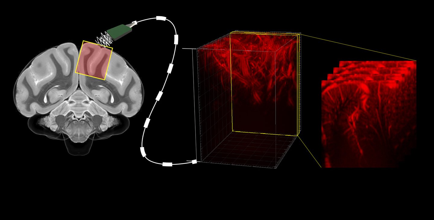
加州理工学院的一项合作开发了一种新型的微创BMI,用于读出与运动计划相对应的大脑活动。
侵入式的脑机接口已经可以让那些因神经损伤或疾病而失去运动能力的人恢复活动能力。不幸的是,只有少数最严重瘫痪的患者有资格并愿意将电极植入他们的大脑。功能性超声(fUS)是一种可以在不损伤脑组织的情况下记录详细的大脑活动的新方法,它可以以100微米的分辨率(单个神经元的大小约为10微米)准确地绘制大脑深处的精确区域的大脑活动。
超声波的工作原理是发射高频声音的脉冲,并测量这些声音振动如何在整个物质(例如人体的各种组织)中回荡。声音以不同的速度穿过这些组织类型,并在它们之间的边界处反射。该技术通常用于拍摄子宫内胎儿的图像,以及用于其他诊断成像。
超声波还可以“听到”器官的内部运动。例如,红细胞,就像一辆经过的救护车一样,当它们接近超声波的来源时,它们的音调会增加,而随着它们流走而减少。测量这种现象使研究人员能够记录大脑血液流动的微小变化,低至100微米(在人类头发宽度的尺度上)。
当大脑的一部分变得更加活跃时,流向该区域的血流量就会增加。功能性超声技术可以产生目标区域中神经信号动力学的详细图像,这是其他非侵入性技术(如fMRI)无法看到的。该技术产生了接近电生理学的细节水平,但侵入性要小得多。
这项技术是在非人类灵长类动物的帮助下开发的,他们被教导做一些简单的任务,包括当出现某些线索时,向某些方向移动眼睛或手臂。当灵长类动物完成任务时,fUS测量后顶叶皮层(PPC)的大脑活动,PPC是大脑中参与计划运动的一个区域。安德森实验室已经研究PPC几十年了,并且以前使用电生理学创建了该区域的大脑活动图。为了验证fUS的准确性,研究人员将fUS的脑成像活动与先前获得的详细电生理学数据进行了比较。
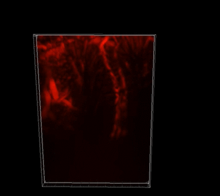
非人灵长类大脑中脉管系统使用功能性超声成像的细节
接下来,通过加州理工学院T&C Chen脑机接口中心的支持,该团队的目标是看看fUS图像中依赖于活动的变化是否可以用来解码非人类灵长类动物的意图,甚至在它发起运动之前。然后,超声成像数据和相应的任务由机器学习算法处理,该算法学习哪些大脑活动模式与哪些任务相关。一旦算法被训练,它就会呈现从非人类灵长类动物那里实时收集的超声波数据。该算法在几秒钟内预测了非人类灵长类动物将要执行的行为(眼球运动或伸展),运动方向(左或右),以及它们计划何时进行运动。
功能性超声成像设法以比功能性MRI高10倍的灵敏度和更高的分辨率记录这些信号。这一发现是基于功能性超声的脑机接口成功的核心。
创新关键点
研究人员使用的功能性超声波(fUS)技术可以以100微米的分辨率(单个神经元的大小约为10微米)准确地绘制大脑深处的精确区域的大脑活动。
创新价值
目前的高分辨率脑机接口使用需要脑部手术的电极阵列,包括打开硬脑膜,颅骨和大脑之间的强纤维膜,并将电极直接植入大脑。但超声波信号可以非侵入性地通过硬脑膜和大脑。只需要在颅骨中植入一个小的超声波透明窗口;这种手术的侵入性明显小于植入电极所需的手术。
新型fUS技术是创建侵入性较小但仍然功能强大的BMI的重要一步。
The new minimally invasive BMI uses fUS technology to accurately map brain activity
A Caltech collaboration has developed a novel minimally invasive BMI for reading out brain activity that corresponds to an exercise program.
Invasive brain-computer interfaces can already restore mobility to people who have lost their ability to move due to neurological damage or disease. Unfortunately, only a few of the most severely paralyzed patients are eligible and willing to have electrodes implanted in their brains. Functional ultrasound (fUS) is a new method that can record detailed brain activity without damaging brain tissue. It can accurately map brain activity in precise regions deep in the brain with a resolution of 100 microns (the size of a single neuron is about 10 microns).
Ultrasound works by sending out pulses of high-frequency sound and measuring how these sound vibrations reverberate through entire substances, such as the body's various tissues. Sound travels through these tissue types at different speeds and bounces off the boundaries between them. The technique is commonly used to take images of fetuses in utero, as well as for other diagnostic imaging.
Ultrasound can also "hear" the internal movements of organs. For example, red blood cells, like a passing ambulance, increase in pitch as they approach the source of the ultrasound and decrease as they flow away. Measuring this phenomenon allowed the researchers to record tiny changes in brain blood flow, as low as 100 microns (on the scale of the width of a human hair).
When a part of the brain becomes more active, blood flow to that area increases. Functional ultrasound technology can produce detailed images of the dynamics of neural signals in the target area that cannot be seen with other non-invasive techniques, such as fMRI. The technique yields a level of detail close to that of electrophysiology, but is much less invasive.
The technique was developed with the help of non-human primates who were taught to do simple tasks, including moving their eyes or arms in certain directions when certain cues were presented. As the primates completed the task, fUS measured brain activity in the posterior parietal cortex (PPC), an area of the brain involved in planning movements. The Anderson lab has been studying PPC for decades and previously used electrophysiology to create a map of brain activity in the region. To verify the accuracy of fUS, the researchers compared fUS brain imaging activity with previously obtained detailed electrophysiological data.
Next, with support from the T&C Chen Center for Brain-Computer Interfaces at Caltech, the team aimed to see if activity-dependent changes in fUS images could be used to decode the intentions of a non-human primate even before it initiates movement. The ultrasound imaging data and corresponding tasks are then processed by machine learning algorithms that learn which brain activity patterns are relevant to which tasks. Once the algorithm is trained, it presents ultrasound data collected in real time from non-human primates.The algorithm predicts within seconds the behavior the non-human primate will perform (eye movement or stretch), the direction of movement (left or right), and when they plan to perform the movement.
Functional ultrasound imaging manages to record these signals with 10 times more sensitivity and higher resolution than functional MRI. This finding is central to the success of brain-computer interfaces based on functional ultrasound.
智能推荐
神经科学创新 | 在基因中创新引入神经保护蛋白可延缓ALS进展
2022-11-17加州大学圣地亚哥分校开发了一种新型基因治疗方法,在家族性渐冻症(ALS)的人化小鼠和大鼠模型中,明显推迟了疾病的发病时间。
涉及学科涉及领域研究方向创新利用数学方法发现食物特异性神经元
2022-08-29麻省理工学院的研究团队研究了人们观看各种图像时的全脑功能性核磁共振成像(fMRI)反应,并创新性地应用了一种数学方法,在FFA两侧的两个神经元簇中发现了一种能够对所有食物产生反应的神经元集群。研究人员将其称之为腹侧食物成分(VFC)。此外,研究团队还开发了一个VFC计算模型,避免了收集fMRI数据的麻烦,还能够用来分析更大的数据集。
涉及学科涉及领域研究方向利用微型硅探头可高清记录大脑活动
2022-08-08由伦敦大学学院的科学家们组成的一个团队开发了一种新的设备,它可以让研究人员以一种前所未有的方式绘制控制行为和决策的复杂神经网络的活动,从而彻底改变我们对大脑的认识。
涉及学科涉及领域研究方向